
The potentiation of MMP-9 promoter activity functionally depends on an upstream peroxisome proliferator-responsive element-like binding site, which displayed an increased DNA binding of a PPAR immunopositive complex. In contrast, the IL-1 -induced DNA-binding of nuclear factor B was significantly impaired by PPAR agonists
All PPARs heterodimerize with the retinoid X receptor (RXR) and bind to specific regions on the DNA of target genes. These DNA sequences are termed PPREs (peroxisome proliferator hormone response elements). The DNA consensus sequence is AGGTCAXAGGTCA, with X being a random nucleotide. In general, this sequence occurs in the promotor region of a gene, and, when the PPAR binds its ligand, transcription of target genes is increased or decreased, depending on the gene. The RXR also forms a heterodimer with a number of other receptors (e.g., vitamin D and thyroid hormone).
The function of PPARs is modified by the precise shape of their ligand-binding domain (see below) induced by ligand binding and by a number of coactivator and corepressor proteins, the presence of which can stimulate or inhibit receptor function, respectively.[7]
Endogenous ligands for the PPARs include free fatty acids and eicosanoids. PPARγ is activated by PGJ2 (a prostaglandin). In contrast, PPARα is activated by leukotriene B4.
Peroxisome proliferator-activated receptor
PPAR -alpha and -gamma pathways.
In the field of molecular biology, the peroxisome proliferator-activated receptors (PPARs) are a group of nuclear receptor proteins that function as transcription factors regulating the expression of genes.[1] PPARs play essential roles in the regulation of cellular differentiation, development, and metabolism (carbohydrate, lipid, and protein) of higher organisms.[2][3]
Nomenclature and tissue distribution
Three types of PPARs have been identified: alpha, gamma, and delta (beta):[2]
α (alpha) - expressed in liver, kidney, heart, muscle, adipose tissue, and others
β/δ (beta/delta) - expressed in many tissues but markedly in brain, adipose tissue, and skin
γ (gamma) - although transcribed by the same gene, this PPAR through alternative splicing is expressed in three forms:
γ1 - expressed in virtually all tissues, including heart, muscle, colon, kidney, pancreas, and spleen
γ2 - expressed mainly in adipose tissue (30 amino acids longer)
γ3 - expressed in macrophages, large intestine, white adipose tissue.
History
PPARs were originally identified in Xenopus frogs as receptors that induce the proliferation of peroxisomes in cells.[4] The first PPAR (PPARα) was discovered during the search of a molecular target for a group of agents then referred to as peroxisome proliferators, as they increased peroxisomal numbers in rodent liver tissue, apart from improving insulin sensitivity.[5] These agents, pharmacologically related to the fibrates were discovered in the early 1980s. When it turned out that PPARs played a much more versatile role in biology, the agents were in turn termed PPAR ligands. The best-known PPAR ligands are the thiazolidinediones; see below for more details.
After PPARδ (delta) was identified in humans in 1992,[6] it turned out to be closely-related to the PPARβ (beta) previously described during the same year in other animals (Xenopus). The name PPARδ is generally used in the US, whereas the use of the PPARβ denomination has remained in Europe where this receptor was initially discovered in Xenopus.
Physiological function
All PPARs heterodimerize with the retinoid X receptor (RXR) and bind to specific regions on the DNA of target genes. These DNA sequences are termed PPREs (peroxisome proliferator hormone response elements). The DNA consensus sequence is AGGTCAXAGGTCA, with X being a random nucleotide. In general, this sequence occurs in the promotor region of a gene, and, when the PPAR binds its ligand, transcription of target genes is increased or decreased, depending on the gene. The RXR also forms a heterodimer with a number of other receptors (e.g., vitamin D and thyroid hormone).
The function of PPARs is modified by the precise shape of their ligand-binding domain (see below) induced by ligand binding and by a number of coactivator and corepressor proteins, the presence of which can stimulate or inhibit receptor function, respectively.[7]
Endogenous ligands for the PPARs include free fatty acids and eicosanoids. PPARγ is activated by PGJ2 (a prostaglandin). In contrast, PPARα is activated by leukotriene B4.
Genetics
The three main forms are transcribed from different genes:
PPARα - chromosome 22q12-13.1 (OMIM 170998)
PPARβ/δ - chromosome 6p21.2-21.1 (OMIM 600409)
PPARγ - chromosome 3p25 (OMIM 601487).
Hereditary disorders of all PPARs have been described, generally leading to a loss in function and concomitant lipodystrophy, insulin resistance, and/or acanthosis nigricans.[8] Of PPARγ, a gain-of-function mutation has been described and studied (Pro12Ala) which decreased the risk of insulin resistance; it is quite prevalent (allele frequency 0.03 - 0.12 in some populations).[9] In contrast, pro115gln is associated with obesity. Some other polymorphisms have high incidence in populations with elevated body mass indexes.
Structure
Like other nuclear receptors, PPARs are modular in structure and contain the following functional domains:
(A/B) N-terminal region
(C) DBD (DNA-binding domain)
(D) flexible hinge region
(E) LBD (ligand binding domain)
(F) C-terminal region
The DBD contains two zinc finger motifs, which bind to specific sequences of DNA known as hormone response elements when the receptor is activated. The LBD has an extensive secondary structure consisting of 13 alpha helices and a beta sheet.[10] Natural and synthetic ligands bind to the LBD, either activating or repressing the receptor.
Pharmacology and PPAR modulators
Main article: PPAR modulator
PPARα and PPARγ are the molecular targets of a number of marketed drugs, e.g. the fibrates. The synthetic chemical perfluorooctanoic acid activates PPARα while the synthetic perfluorononanoic acid activates both PPARα and PPARγ.
See also
Thiazolidinedione
Anti-diabetic drug
Diabetes mellitus
Insulin resistance
Metabolic syndrome
References
^ Michalik L, Auwerx J, Berger JP, Chatterjee VK, Glass CK, Gonzalez FJ, Grimaldi PA, Kadowaki T, Lazar MA, O'Rahilly S, Palmer CN, Plutzky J, Reddy JK, Spiegelman BM, Staels B, Wahli W (2006). "International Union of Pharmacology. LXI. Peroxisome proliferator-activated receptors". Pharmacol. Rev. 58 (4): 726–41. doi:10.1124/pr.58.4.5. PMID 17132851.
^ a b Berger J, Moller DE (2002). "The mechanisms of action of PPARs". Annu. Rev. Med. 53: 409–35. doi:10.1146/annurev.med.53.082901.104018. PMID 11818483.
^ Feige JN, Gelman L, Michalik L, Desvergne B, Wahli W (2006). "From molecular action to physiological outputs: peroxisome proliferator-activated receptors are nuclear receptors at the crossroads of key cellular functions". Prog. Lipid Res. 45 (2): 120–59. doi:10.1016/j.plipres.2005.12.002. PMID 16476485.
^ Dreyer C, Krey G, Keller H, Givel F, Helftenbein G, Wahli W (1992). "Control of the peroxisomal beta-oxidation pathway by a novel family of nuclear hormone receptors". Cell 68 (5): 879–87. doi:10.1016/0092-8674(92)90031-7. PMID 1312391.
^ Issemann I, Green S (1990). "Activation of a member of the steroid hormone receptor superfamily by peroxisome proliferators". Nature 347 (6294): 645–50. doi:10.1038/347645a0. PMID 2129546.
^ Schmidt A, Endo N, Rutledge SJ, Vogel R, Shinar D, Rodan GA (1992). "Identification of a new member of the steroid hormone receptor superfamily that is activated by a peroxisome proliferator and fatty acids". Mol. Endocrinol. 6 (10): 1634–41. doi:10.1210/me.6.10.1634. PMID 1333051.
^ Yu S, Reddy JK (2007). "Transcription coactivators for peroxisome proliferator-activated receptors". Biochim. Biophys. Acta 1771 (8): 936–51. doi:10.1016/j.bbalip.2007.01.008. PMID 17306620.
^ Meirhaeghe A, Amouyel P (2004). "Impact of genetic variation of PPARgamma in humans". Mol. Genet. Metab. 83 (1-2): 93–102. doi:10.1016/j.ymgme.2004.08.014. PMID 15464424.
^ Buzzetti R, Petrone A, Ribaudo MC, Alemanno I, Zavarella S, Mein CA, Maiani F, Tiberti C, Baroni MG, Vecci E, Arca M, Leonetti F, Di Mario U (2004). "The common PPAR-gamma2 Pro12Ala variant is associated with greater insulin sensitivity". Eur. J. Hum. Genet. 12 (12): 1050–4. doi:10.1038/sj.ejhg.5201283. PMID 15367918.
^ Zoete V, Grosdidier A, Michielin O (2007). "Peroxisome proliferator-activated receptor structures: ligand specificity, molecular switch and interactions with regulators". Biochim. Biophys. Acta 1771 (8): 915–25. doi:10.1016/j.bbalip.2007.01.007. PMID 17317294.
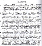
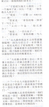
Rat renal mesangial cells express high levels of matrix metalloproteinase 9 (MMP-9) in response to inflammatory cytokines such as interleukin 1 (IL-1 ). We tested whether ligands of the peroxisome proliferator-activated receptor (PPAR ) could influence the cytokine-induced expression of MMP-9.
Different PPAR agonists dose-dependently inhibited the IL-1 -triggered increase in gelatinolytic activity mainly by decreasing the MMP-9 steady-state mRNA levels.
PPAR agonists on their own had no effects on MMP-9 mRNA levels and gelatinolytic activity.
Surprisingly, the reduction of MMP-9 mRNA levels by PPAR activators contrasted with an amplification of cytokine-mediated MMP-9 gene promoter activity and mRNA expression. The potentiation of MMP-9 promoter activity functionally depends on an upstream peroxisome proliferator-responsive element-like binding site, which displayed an increased DNA binding of a PPAR immunopositive complex. In contrast, the IL-1 -induced DNA-binding of nuclear factor B was significantly impaired by PPAR agonists. Most interestingly, in the presence of an inducible nitric-oxide synthase (iNOS) inhibitor, the PPAR -mediated suppression switched to a strong amplification of IL-1 -triggered MMP-9 mRNA expression. Concomitantly, activators of PPAR potentiated the cytokine-induced iNOS expression. Using actinomycin D, we found that NO, but not PPAR activators, strongly reduced the stability of MMP-9 mRNA. In contrast, the stability of MMP-9 protein was not affected by PPAR activators. In summary, our data suggest that the inhibitory effects of PPAR agonists on cytokine-induced MMP-9 expression are indirect and primarily due to a superinduction of iNOS with high levels of NO reducing the half-life of MMP-9 mRNA.
Subscribe to:
Post Comments (Atom)


No comments:
Post a Comment